【临床病史】:患者,12岁男孩,既往健康,在一次癫痫发作后被送入急诊科。12-year-old boy, previously healthy, is brought to the emergency room after an episode of seizures.
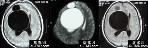
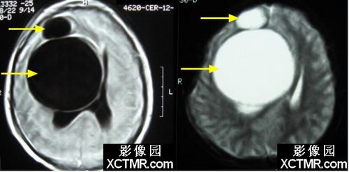
【影像图片】MRI图像

【讨论】:Hydatid disease is a worldwide zoonosis produced by the larval stage of the Echinococcus tapeworm. The two main types of hydatid disease are caused by E granulosus and E multilocularis. E granulosus is the more common type, whereas E multilocularis is less common but more invasive, mimicking a malignancy. It is commonly seen in the great grazing regions of the World, particularly the Mediterranean region, Africa, South America, the Middle East, Australia, and New Zealand.包虫病是一种流行于全世界范围的动物源性寄生虫病,主要是由棘虫绦虫幼虫期所引发。引发包虫病的两种主要的寄生虫类型分别是细粒棘球绦虫和多房棘球绦虫,细粒棘球绦虫更常见,多房棘球绦虫更少见但侵袭性更强,其表现类似于恶性病变。它常见于世界上的的牧区,特别是地中海区域、非洲、南美、东亚、澳大利亚和新西
Dogs or other carnivores are definitive hosts, whereas sheep or other ruminants are intermediate hosts. Humans are secondarily infected by the ingestion of food or water that has been contaminated by dog feces containing the eggs of the parasite.狗或其他的肉身动物是终宿主,而羊或其他的反刍性动物是中间宿主。被包含有寄生虫卵的狗粪所污染的食物或水被人类摄入从而引起继发性感染。
Intracranial granulosus echinococcosis occurs in only approximately 2% of cases of hydatid disease, typically involving the cerebral parenchyma, especially the parietal lobes, corresponding to the middle cerebral artery watershed territory. Intracranial subarachnoid spaces are the second most common location of the disease in the CNS, although their occurrence is far less frequent. Cases of cerebral aqueduct cyst, gigantic cyst arising from the diploe of cranial bones with intracranial extension, and intradural spinal hydatid cysts have been reported. Cysts are usually single and may be unilocular or multilocular. Cerebral hydatid cyst is more common in children than in adults.颅内的细粒棘球绦虫感染仅见于约2%的包虫病病例,通常累及大脑实质,特别是顶叶,符合大脑中动脉分水岭区,虽然很少见,但颅内的蛛网膜下腔是第二常见的发病部位。发生于大脑导水管的囊肿、起源于颅骨板障并延伸至颅内的巨大囊肿、以及椎管内硬膜下囊肿都有报道。囊肿常常是单发的,可以是单房或多房。儿童大脑包虫囊肿比成人更常见。
At MRI, cerebral hydatid disease generally appears unilocular and is isointense relative to cerebrospinal fluid. The lack of surrounding edema and the marked mass effect make it easy to distinguish cerebral hydatid disease from abscess and cystic tumor. The presence of a hypointense rim, especially on T2-weighted MR images, is characteristic of hydatid cyst of the brain. Cerebral hydatid cyst is generally solitary but may be multiple when it ruptures spontaneously or due to trauma or surgery. Multivesicular cysts are rare in the brain. Calcification occurs in less than 1% of cases.在MRI上,大脑包虫病通常表现为单房病变,信号与脑脊液相仿。无周边水肿,明显的占位效应可以与脓肿和其他囊性肿瘤相鉴别。病变可以出现一个低信号环,尤其是在T2序列上,这是脑包虫囊肿特征性病变。颅内的多囊状病变是相当少的,不超过1%的病例可以出现钙化。
转自——影像园